
From Blood Draw to PRP
Follow one platelet from the patient’s bloodstream to the final PRP syringe.
What changes between the blood draw and the final syringe?
Lesson 1 introduced the platelet. Now follow the whole sample and see how the preparation process changes where blood components are located and which fraction is ultimately collected.
Everything begins in whole blood
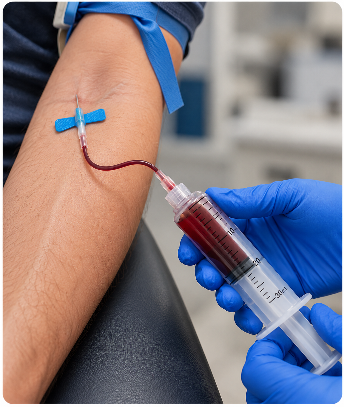
The starting sample contains the full blood mixture.
Red blood cells, white blood cells, plasma, and platelets are together in the patient’s circulating blood. The collection method varies by PRP system, but the preparation always begins with the patient’s own blood.
Blood is designed to clot. PRP preparation needs time.
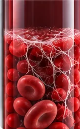 Before anticoagulation
Instructions: Select Add citrate to the sample to see how anticoagulation helps keep blood processable.
For many conventional injectable PRP systems, blood is collected with a citrate-based anticoagulant such as ACD-A or sodium citrate. Citrate binds available calcium and interrupts calcium-dependent steps in coagulation, helping keep the sample processable while the PRP is prepared.
Calcium is required for normal coagulation. Citrate chelates available calcium, reducing calcium-dependent clotting activity while the sample is being processed.
Anticoagulant choice, amount, and use vary by device and protocol. Staff should follow the instructions for the specific system being used.The container is part of the process
PRP is not always prepared in a simple straight tube. Vessel geometry and collection design can influence where separated components go and what can ultimately be recovered.
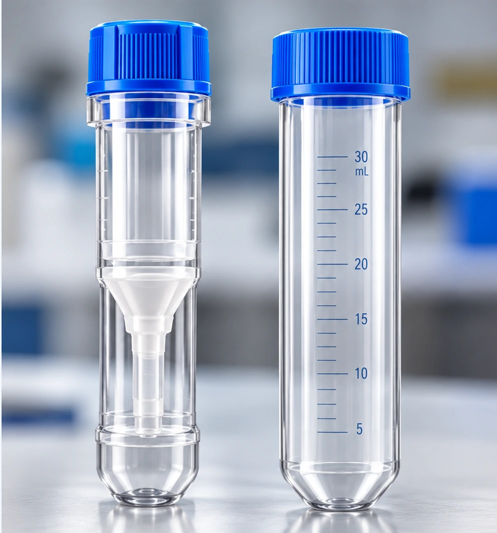
Select each of the four features below to explore how the vessel affects PRP preparation.
Explore all four to see why the vessel is more than a container.
Want to go a little deeper? Select the section below to learn why vessel geometry can influence PRP separation.
Vessel shape changes the path components travel during centrifugation and can change which regions are accessible afterward. Sample volume, barriers, constrictions, and collection ports can also influence what is ultimately recovered.
The centrifuge changes where components are located
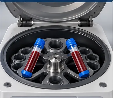
Centrifugation exposes the sample to force far greater than normal gravity. Blood components move differently according to their sedimentation behavior, while the vessel, rotor, force, time, and protocol influence the separation.
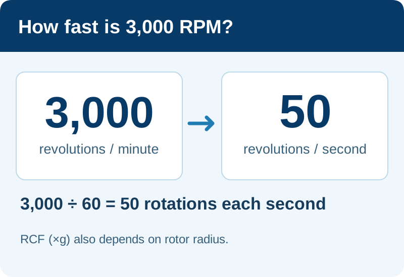
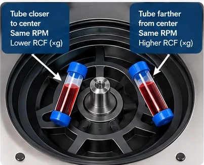
Picture the centrifuge rotor. At an illustrative speed of 3,000 RPM, it makes 50 complete revolutions every second (3,000 ÷ 60). At the same RPM, a sample farther from the rotor axis experiences greater relative centrifugal acceleration.
RPM (revolutions per minute) tells you how fast the rotor turns. RCF (relative centrifugal force), expressed as ×g, describes the force experienced by the sample and accounts for rotor radius, making it more useful when comparing centrifugation conditions.
Same sample. New geography.
Before centrifugation the components are mixed. After centrifugation, visibly different regions emerge.
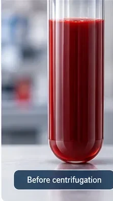
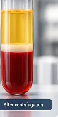
Red blood cells have moved toward the more sedimented region, while plasma remains above. A thin pale interface can contain many white blood cells (leukocytes) and platelets. But that interface is not a universal, perfectly sharp “PRP layer.”
Platelets are not necessarily sitting in one visible stripe
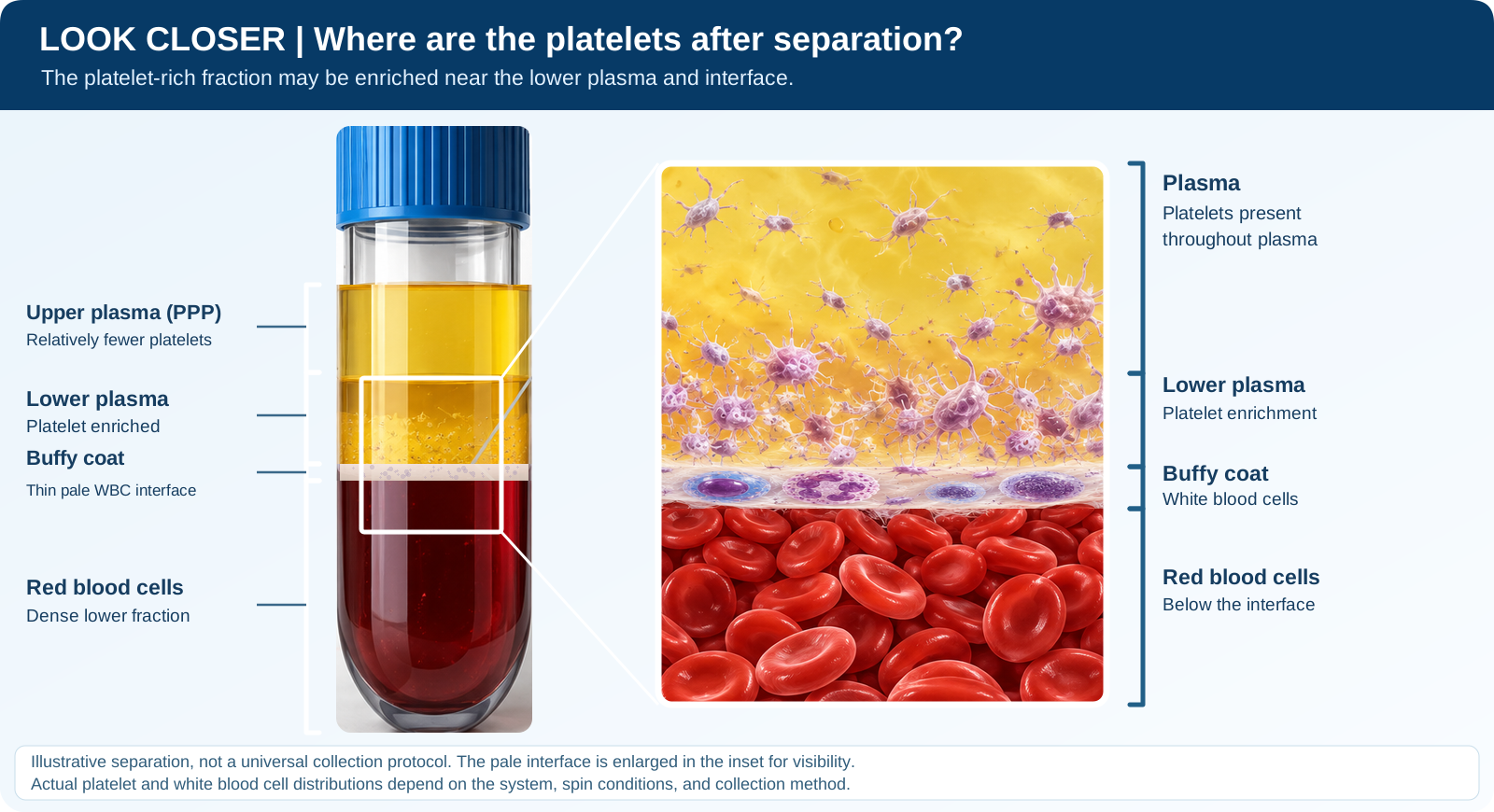
Select all four components below to explore the separated sample.
Click all four components. The important lesson is that what you can see macroscopically does not tell you exactly where every platelet is.
Some platelets in this illustration have an activated appearance, with projections extending from their surfaces. Resting platelets typically have a smoother, disc-like shape.
Platelets may become activated during collection or processing, depending on the preparation method and handling conditions. However, not every PRP preparation contains the same proportion of activated platelets.
This illustration is intended to show where platelets may be distributed after separation, not to represent the activation state of every platelet in a PRP preparation.
PPP does not necessarily mean “platelet-free”
PPP stands for Platelet-Poor Plasma. Which description is most accurate?
What you collect helps determine what you get
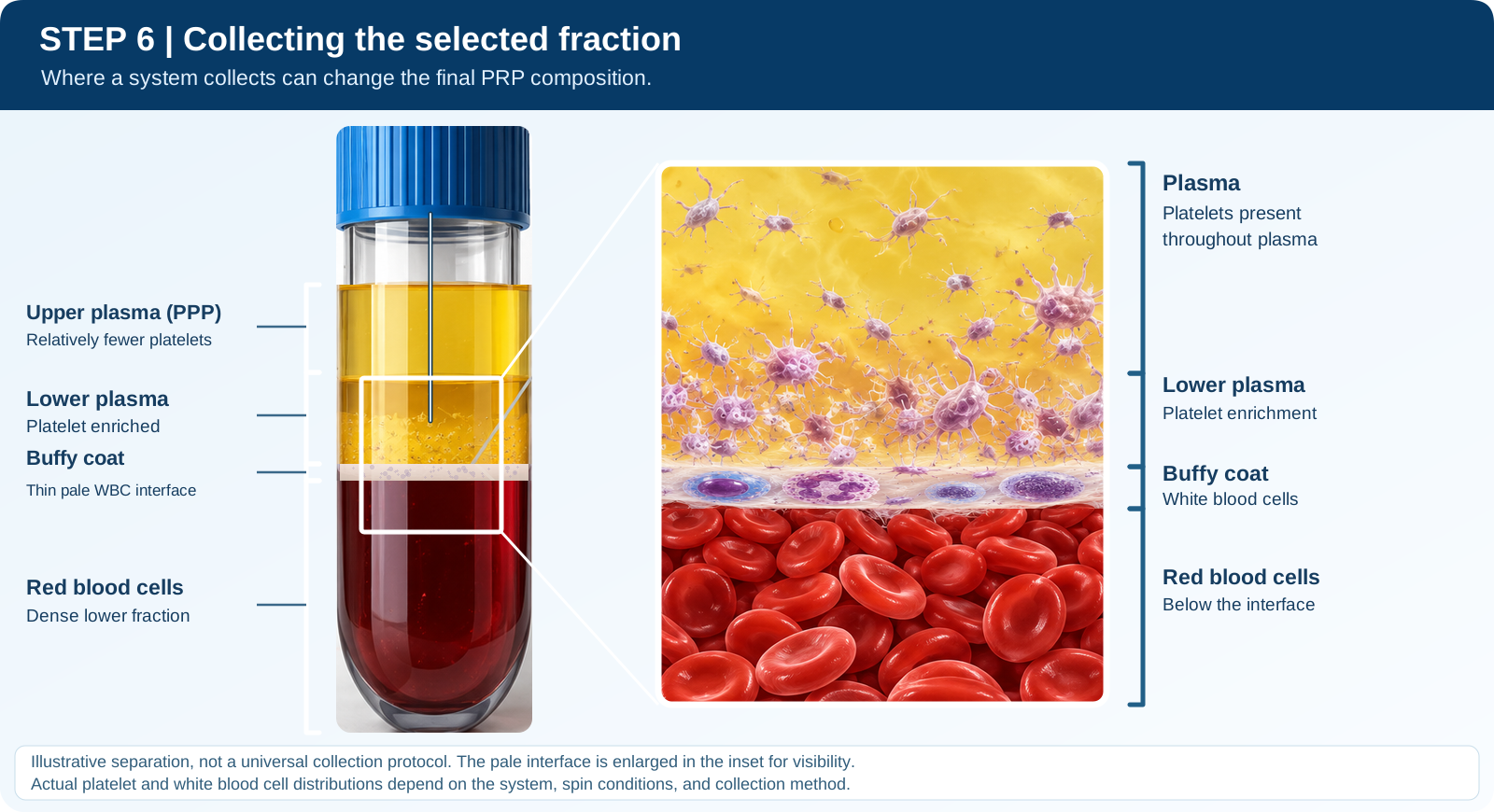
Different systems are designed to recover different portions of the separated sample. Explore three collection concepts. These are tendencies, not universal numerical recipes.
Select each of the three collection options below to see how the final PRP can differ.
The system and protocol determine which fraction becomes the final preparation.
Most of the starting blood does not end up in the PRP syringe
If the final PRP volume is only a few milliliters, where did the rest of the starting sample go?
Same starting blood does not guarantee the same final PRP
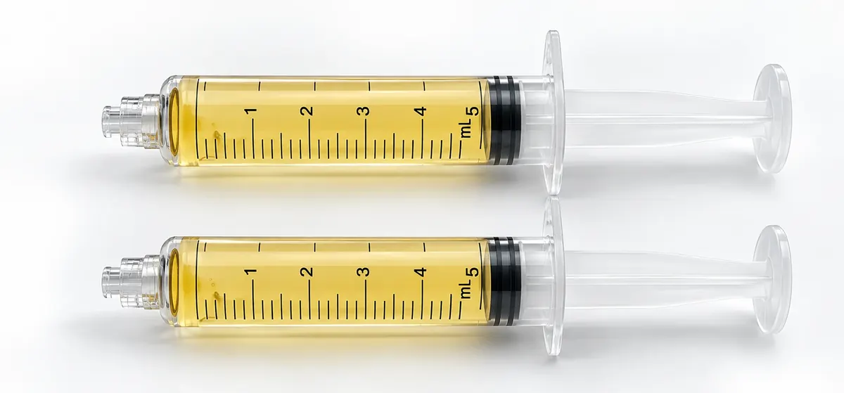
Must the two final preparations be biologically identical?
Even when the same patient receives PRP using the same preparation system, platelet counts and other blood components may vary between visits. The final PRP preparation may therefore differ, too.
Same patient. Same system. Different day. Potentially different PRP. Read the study.Turn the process into a patient-friendly answer
“What happens to my blood after you draw it?”
Understanding variability does not mean declaring one system “best”
“Which PRP system gives the best PRP?”
Know it. Explain it. Know when to escalate it.
PRP begins as whole blood. Anticoagulation when required, separator design, centrifugation conditions, separation, and collection all influence what reaches the final syringe.
PRP is a living, biologically active preparation. Over time, platelets and other blood components can undergo changes that may affect the characteristics of the final product.
Many PRP systems specify a maximum four-hour window for use, although the exact timing and handling requirements vary by manufacturer.
Best practice: Prepare and use PRP as promptly as practical, following the manufacturer’s instructions for use. A four-hour window does not mean the preparation remains biologically unchanged throughout that time.
Now that you know how the sample is separated and collected, we can ask the next question: exactly what did we collect?
Lesson 2 intentionally teaches preparation concepts rather than a universal PRP recipe. Published literature documents substantial variation in anticoagulants, vessels, centrifugation methods, collection strategies, and final PRP composition.
Show that you understand the concepts
This is not a trick quiz. The questions are designed to confirm that you can apply the lesson, not simply recognize a definition. Pass this lesson check with 80%. The final course assessment will require 90% for the certificate.