PRPdose Clinical Science

The Cellular Conversation

The centrifuge creates the cellular gradient. The collection strategy shapes which cells become part of the final preparation and the biological conversation that follows.

Platelet Monocyte Neutrophil Red blood cell
Continue to the Science 01 · CREATE THE GRADIENT Leukocyte-Poor Collect above the interface Monocyte-Retaining Enter the upper buffy coat Leukocyte-Rich Reach the lower buffy coat LP-PRP MR / NP-PRP LR-PRP LP-PRP Platelet-centered signaling Lower leukocyte environment MR / NP-PRP Platelet-monocyte crosstalk Resolving macrophage functions LR-PRP Broader leukocyte signaling Stronger innate immune activity THE CENTRAL IDEA The centrifuge creates the gradient. The clinician defines the conversation. The biology begins with what is intentionally collected. The centrifuge creates a cellular density gradient.
Chapter 1 · Composition
Same platelets. Different supporting cells.

Three preparations can create three different signaling environments.

PRP should not be defined by platelet concentration alone. Leukocyte composition, activation state, tissue context, dose, and processing technique all influence the conversation.

Monocyte-Selective PRP

Immune-modulating pathway
Platelets + monocytes · neutrophils limited
Platelet–monocyte crosstalk
Macrophage polarization toward resolving, M2-like functions
Angiogenesis · matrix regulation · organized remodeling
Potential conversationA coordinated inflammatory response that progresses toward resolution and regenerative tissue-appropriate repair.
Leukocyte-Poor PRP

Lower-leukocyte pathway
Platelets · minimal leukocyte recovery
Platelet activation and growth-factor release
Resident chondrocytes, fibroblasts and progenitor cells
Lower leukocyte-associated inflammatory signaling
Potential conversationA comparatively quiet platelet-centered signal that may be desirable in selected intra-articular applications.

Neutrophil-Rich PRP

Innate inflammatory pathway
Platelets + monocytes + greater neutrophil recovery
ROS · proteases · NET-associated signaling
Greater inflammatory and matrix-degrading pressure
Potentially prolonged M1-like signaling in susceptible tissues
Potential conversationA stronger innate immune response that may be useful in some settings but undesirable in others.
Scientific context: These pathways are educational models, not fixed biological outcomes. Macrophages occupy a spectrum of functional states, and neutrophils can be beneficial or detrimental depending on timing, concentration, tissue, indication, and local disease environment.
From biology to hardware

Can your PRP system control the conversation?

The separation device, centrifugation stability, draw-volume capacity, recovery efficiency, and collection method all influence which cells reach the final preparation.

1
Intended biology
Processing system
Final product
Audit Your PRP System →
Chapter 2 · Quantity
If the conversation matters...

How many platelets should participate?

Several authors have proposed platelet dose targets in the range of 4 to 10 billion platelets for selected applications, while optimal dosing remains an active area of investigation.

4–10billion platelets

Concentration is not the same as dose.

A high concentration factor can still produce a modest total dose when the starting blood volume is small. Absolute platelet dose depends on the patient’s baseline platelet count, whole-blood draw volume, system recovery, and final processing losses.

Baseline CBCAvailable platelet inventory
Draw VolumeTotal cells collected
RecoveryCells retained after processing
The deeper question: Reaching a dose target is foundational, but the final product should also be evaluated for cellular composition and quality.
Clinical Reference: We highly recommend referencing the 2026 AAPM&R Guidance Statement on Platelet-Rich Plasma for Knee Osteoarthritis for comprehensive, peer-reviewed clinical protocols and consensus standards.
Chapter 3 · Platelet quality
Are all platelets biologically identical?

The count does not fully describe the cellular payload.

Standard CBC systems quantify platelet number. They generally do not fully characterize platelet maturity, activation state, metabolic activity, or functional heterogeneity.

Young and old platelets may carry different signaling capacity.

Emerging hematology research suggests that immature or reticulated platelets may have greater RNA content, metabolic activity, and granule signaling capacity than older circulating platelets. Mean platelet volume and immature platelet fraction can provide additional context, although these measures are not routinely available in many regenerative medicine offices.

A platelet count tells you how many. It does not automatically tell you which platelet subpopulations were recovered or how functionally active they are.
Illustrative reticulated plateletLarger · RNA-rich · found closer to buffy coat · metabolically active
Older plateletSmaller · found higher above buffy coat · reduced cellular content
Chapter 4 · Patient inventory
How much biology does this patient have available?

The baseline CBC sets the limits of the preparation.

Every patient begins with a different platelet inventory. Precision dosing starts before the centrifuge spins.

Small draws create mathematical ceilings.

A patient with a baseline platelet count of 200 M/µL contributes approximately 3.4 billion platelets to a 20 mL sample before processing losses, assuming roughly 17 mL of actual whole blood after anticoagulant volume.

Starting inventory → processing losses → final injectable dose

Tailor the draw to the intended dose.

Depending on the baseline platelet count, recovery efficiency, and treatment objective, a larger collection may be required. Personalized draw volume allows dose to be planned rather than assumed.

Model the Patient’s Dose →
Chapter 5 · Separation physics
Where do the cells that shape the conversation reside?

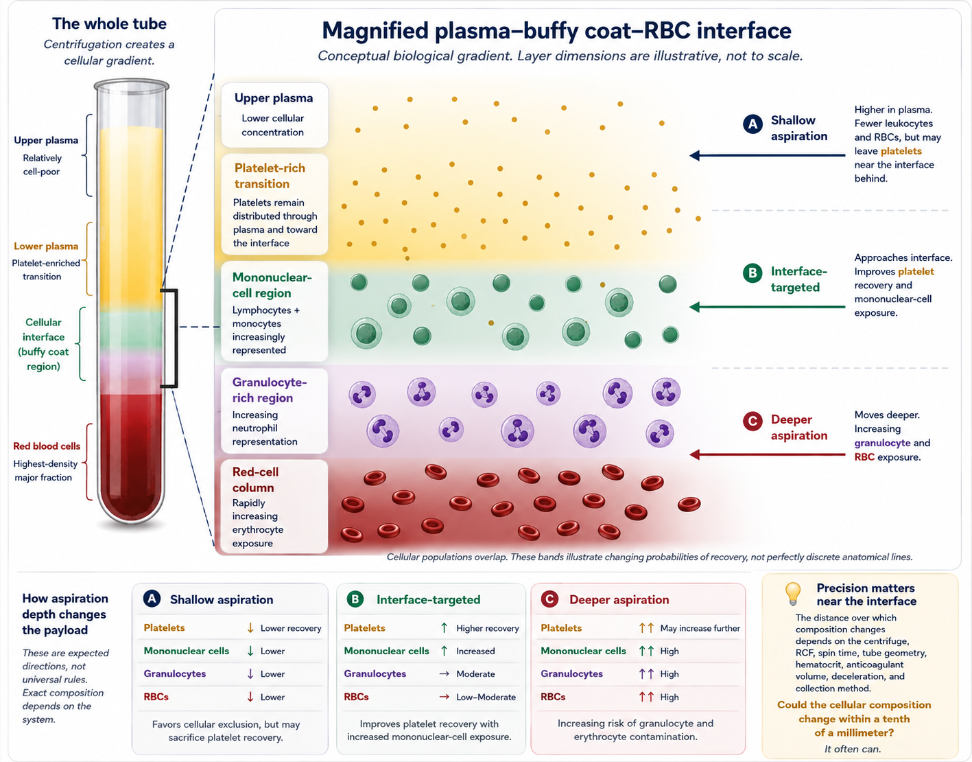
The cellular gradient is a map, not a single line.

Density, size, shape, centrifugal force, spin duration, vessel geometry, and deceleration all influence where cellular populations stratify.

Upper plasmalower cellular content
Lower plasmaplatelet-enriched region
Platelets + mononuclear cellsNeutrophils
Red blood cells
PRPdose “Golden Zone”Lower plasma + upper buffy coatBuffy CoatThin white cellular interface

The conversation can change within a tenth of a millimeter.

The buffy coat is the white interface between plasma and red blood cells. Its upper portion may contain platelets and mononuclear cells, while its deeper portion is generally more granulocyte rich. The PRPdose “Golden Zone” spans the lower plasma and upper buffy coat; moving deeper may increase neutrophil and red-cell recovery, while staying too high may sacrifice platelet recovery and monocytes.

Platelets: distributed through plasma with increasing concentration toward the interface.
Mononuclear cells: often concentrated near the upper interface.
Neutrophils: generally extend deeper into the buffy coat and granulocyte-rich region.
RBCs: should be carefully controlled, particularly for intra-articular preparations.
Chapter 6 · Tissue response
What does an organized conversation build?

The desired matrix depends on the tissue being repaired.

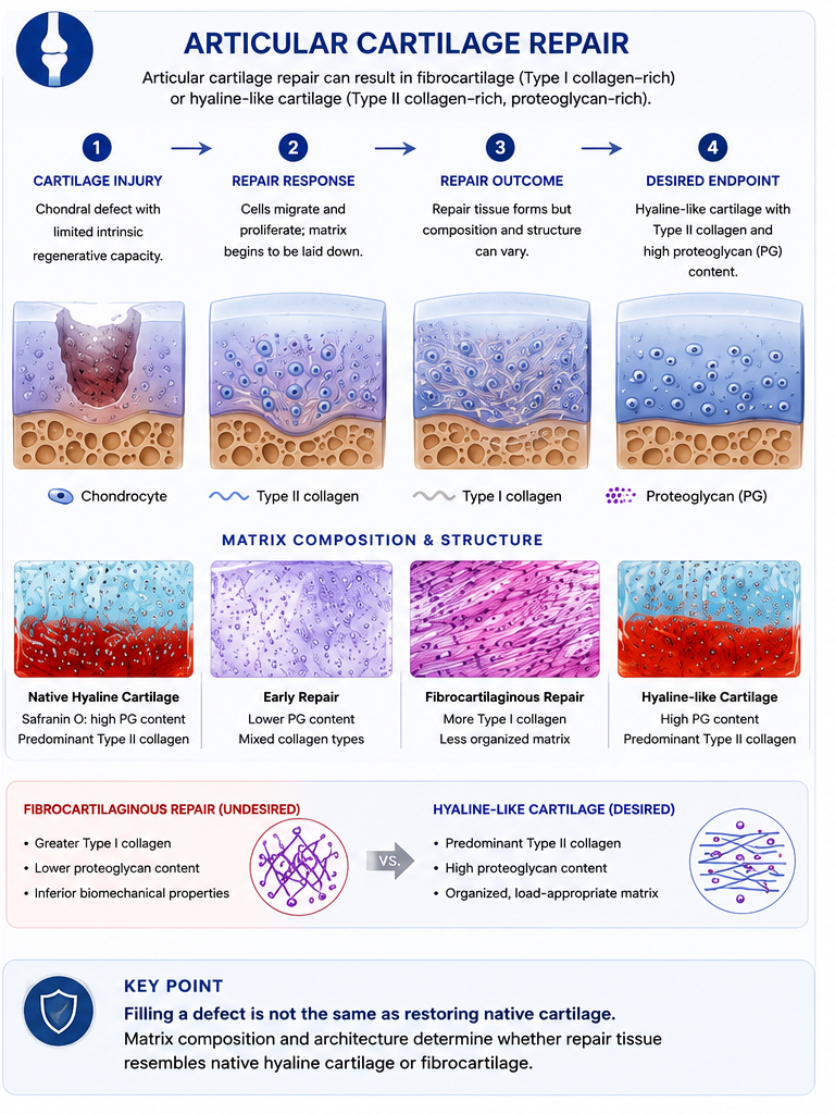
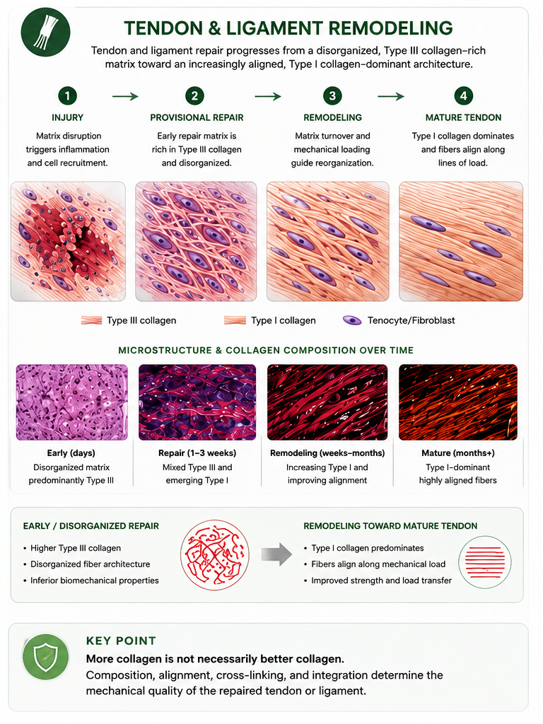
Macrophage signaling and extracellular-matrix remodeling occur before the collagen endpoint. The ideal collagen architecture is not the same in tendon and articular cartilage.

More organized remodeling

Illustrative tissue-specific outcomes when inflammation progresses toward resolution.

Tendon and ligament: aligned Type I collagenEarly Type III-rich matrix remodels toward stronger, load-oriented Type I fibers.
Articular cartilage: Type II-rich hyaline matrixA fine Type II network with proteoglycans supports compression and low-friction motion.

Less mature or disorganized remodeling

Illustrative patterns associated with persistent inflammatory and proteolytic pressure.

Tendon and ligament: disorganized scar tissuePersistent Type III-rich, poorly aligned collagen may contribute to mechanically inferior scar tissue.
Cartilage defect: Type I-rich fibrocartilageIt may fill a defect but does not fully reproduce native Type II-rich articular cartilage.
Put the science into practice

Measure the dose. Evaluate the system. Understand the conversation.

Platelet quantity and cellular composition answer different questions. The next step is to test both the mathematics and the hardware.

Quantify the dose

Use the patient’s baseline CBC, draw volume, and processing recovery to estimate the total platelet dose available for treatment.

Open PRPdose Calculator →

Evaluate the hardware

Review recovery efficiency, RBC exclusion, cellular selectivity, centrifuge stability, draw-volume capability, and workflow repeatability.

Open the PRP System Audit →

Selected scientific foundations

  1. De Matthaeis A, et al. High-Dose Neutrophil-Depleted Platelet-Rich Plasma Therapy for Knee Osteoarthritis (2024).
  2. Corsini A, et al. Re-Evaluating Platelet-Rich Plasma Dosing Strategies in Sports Medicine (2025).
  3. Bansal H, et al. PRP in knee osteoarthritis: correct dose critical for long-term clinical efficacy (2021).
  4. Handtke S, Thiele T. Large and small platelets: (When) do they differ? (2020).
  5. Koupenova M, et al. Circulating Platelets as Mediators of Immunity, Inflammation, and Thrombosis (2018).
Educational and scientific reference only. The technical data, cellular-processing concepts, dose ranges, and proposed biological pathways presented on this page are intended exclusively for licensed healthcare professionals, researchers, and industry evaluation. This material is educational and scientific reference content only. It is not medical advice, does not prescribe a treatment protocol, and does not guarantee that any specific PRP system or preparation will produce a particular cellular response or clinical outcome. Macrophage states, leukocyte effects, platelet biology, and tissue remodeling vary with patient factors, preparation method, dose, timing, tissue, and indication. PRP devices are regulated for specific cleared indications, and broader clinical applications may be investigational or off-label. Clinicians must use independent clinical judgment and verify decisions against primary literature, manufacturer instructions, institutional policy, and applicable regulation.